Lineaging C. elegans Embyonic Development.
Our system allows us to reconstruct the lineage of a C. elegans embryo with minimal manual annotation. The three movies below show the complexity of worm development and our ability to resolve and track cell identity. The last figure is an image of a cell lineage map we have constructed from our data.
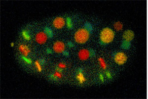
A Depth-Coded Movie of the Development of a Worm.
In this movie a worm develops from four cells (ABa, ABp, EMS, and P1) to a hatched larva. Each time point represents a 3D image of the embryo, with the nuclei artificially colored to reflect their depth (red is closest and blue is farthest away).
This file is 20 MB and takes quite awhile to load.
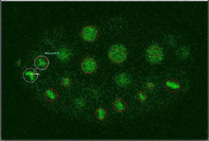
A Movie Tracking the Anterior-Most Daughter of Progressive Cell Divisions.
Only a single plane centered on the cell of interest is shown for each time point. The tracked cell is surrounded by a white circle, while others are shown surrounded by a red circle. The sister of a newly divided cell is shown connected by a line for one frame.
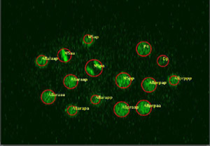
A Movie Showing Each Nucleus in an Embryo at One Time.
This movie illustrates serial optical sections through an embryo at one time point during development, moving from top to bottom. MSaa is at metaphase of the cell cycle. MSpa is at anaphase.
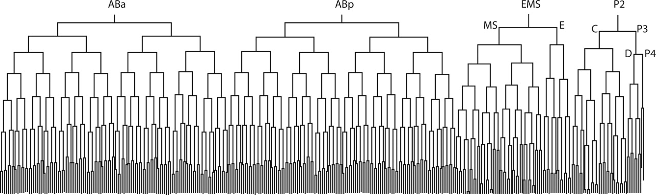
The Embryonic Lineage of C. elegans
This image of the lineage of an embryo past the 350-cell stage was generated by StarryNite and AceTree.